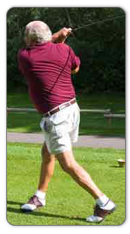
Shoe Lifts For Leg Length Discrepancy
There are not one but two different types of leg length discrepancies, congenital and acquired. Congenital means that you are born with it. One leg is structurally shorter compared to the other. As a result of developmental stages of aging, the brain senses the gait pattern and identifies some variation. The human body typically adapts by dipping one shoulder over to the "short" side. A difference of under a quarter inch isn't grossly uncommon, demand Shoe Lifts to compensate and generally does not have a profound effect over a lifetime.

Leg length inequality goes typically undiagnosed on a daily basis, however this condition is easily solved, and can eradicate many cases of lower back pain.
Therapy for leg length inequality commonly involves Shoe Lifts . These are generally affordable, frequently priced at under twenty dollars, in comparison to a custom orthotic of $200 plus. Differences over a quarter inch can take their toll on the spine and should probably be compensated for with a heel lift. In some cases, the shortage can be so extreme that it requires a full lift to both the heel and sole of the shoe.
Chronic back pain is the most common condition impacting men and women today. Over 80 million men and women suffer from back pain at some stage in their life. It is a problem that costs businesses millions yearly because of lost time and output. Fresh and improved treatment methods are constantly sought after in the hope of minimizing the economical influence this condition causes.

People from all corners of the earth suffer from foot ache as a result of leg length discrepancy. In these situations Shoe Lifts can be of very beneficial. The lifts are capable of alleviating any pain and discomfort in the feet. Shoe Lifts are recommended by numerous specialist orthopaedic physicians.
In order to support the human body in a well-balanced fashion, the feet have a very important role to play. In spite of that, it is sometimes the most overlooked zone of the body. Many people have flat-feet which means there is unequal force exerted on the feet. This will cause other parts of the body including knees, ankles and backs to be impacted too. Shoe Lifts guarantee that proper posture and balance are restored.
Leg length inequality goes typically undiagnosed on a daily basis, however this condition is easily solved, and can eradicate many cases of lower back pain.
Therapy for leg length inequality commonly involves Shoe Lifts . These are generally affordable, frequently priced at under twenty dollars, in comparison to a custom orthotic of $200 plus. Differences over a quarter inch can take their toll on the spine and should probably be compensated for with a heel lift. In some cases, the shortage can be so extreme that it requires a full lift to both the heel and sole of the shoe.
Chronic back pain is the most common condition impacting men and women today. Over 80 million men and women suffer from back pain at some stage in their life. It is a problem that costs businesses millions yearly because of lost time and output. Fresh and improved treatment methods are constantly sought after in the hope of minimizing the economical influence this condition causes.
People from all corners of the earth suffer from foot ache as a result of leg length discrepancy. In these situations Shoe Lifts can be of very beneficial. The lifts are capable of alleviating any pain and discomfort in the feet. Shoe Lifts are recommended by numerous specialist orthopaedic physicians.
In order to support the human body in a well-balanced fashion, the feet have a very important role to play. In spite of that, it is sometimes the most overlooked zone of the body. Many people have flat-feet which means there is unequal force exerted on the feet. This will cause other parts of the body including knees, ankles and backs to be impacted too. Shoe Lifts guarantee that proper posture and balance are restored.
Hammer Toe Fusion Surgery
The term hammertoes describes three unique contracture deformities of the toes. The deformities differ by the location of contracture in each joint of the toe. The three deformities include hammer toe, claw toe and mallet toe. Hammer toes may be flexible or rigid. Hammer toes are most common on the lesser toes (2-5) and may affect one or more toes simultaneously. Hallux malleus is the term used to described a hammer toe of the great toe. Hallux malleus is often found as an isolated foot problem. Hammer toes are found equally in men and women. The onset of hammer toes is between the ages of 30 and 80 years of age.
Causes
Essentially, hammertoes are caused by an abnormal interworking of the bones, muscles, ligaments and tendons that comprise your feet. When muscles fail to work in a balanced manner, the toe joints can bend to form the hammertoe shape. If they remain in this position for an extended period, the muscles and tendons supporting them tighten and remain in that position. A common factor in development of hammertoe is wearing shoes that squeeze the toes or high heels that jam the toes into the front of the shoe. Most likely due to these factors, hammertoe occurs much more frequently in women than in men.
 Symptoms
A toe stuck in an upside-down "V" is probably a hammertoe. Some symptoms are, pain at the top of the bent toe when putting on a shoe. Corns forming on the top of the toe joint. The toe joint swelling and taking on an angry red colour. Difficulty in moving the toe joint and pain when you try to so. Pain on the ball of the foot under the bent toe. Seek medical advice if your feet regularly hurt, you should see a doctor or podiatrist. If you have a hammertoe, you probably need medical attention. Ask your doctor for a referral to a podiatrist or foot surgeon. Act now, before the problem gets worse.
Diagnosis
First push up on the bottom of the metatarsal head associated with the affected toe and see if the toe straightens out. If it does, then an orthotic could correct the problem, usually with a metatarsal pad. If the toe does not straighten out when the metatarsal head is pushed up, then that indicates that contracture in the capsule and ligaments (capsule contracts because the joint was in the wrong hammertoes position for too long) of the MTP joint has set in and surgery is required. Orthotics are generally required post-surgically.
Non Surgical Treatment
Inserts in your shoes can be used to help relieve pressure on the toes from the deformity. Splints/Straps. These can be used to help re-align and stretch your toes and correct the muscle imbalance and tendon shortening. One of the most common types are toe stretchers like the yogatoe. Chiropody. A chiropodist can remove calluses or corns, areas of hard skin that have formed to make the foot more comfortable.Steroid injections can help to reduce pain and inflammation.
Surgical Treatment
Until recently, wires were used for surgical correction. In this technique, one or more wires are inserted into the bone through both the affected joint and a normally healthy toe joint, and the end of the toe. These wires stay in place for four to six weeks, protruding from the end of the toes. Due to the protruding wire, simple things such working, driving, bathing and even sleeping are difficult while these wires are in place. During this recovery period, patients often experience discomfort during sleep and are subject possible infection.
 Prevention
Have your feet properly measured, make sure that, while standing, there is a centimetre (? thumb) of space for your longest toe at the end of each shoe. Buy shoes that fit the longer foot. Shop at the end of the day, when foot swelling is greatest. Don't go by numbers, sizes vary by brand, so make certain your shoes are comfortable. Wear wide shoes with resilient soles, avoid shoes with pointed toes.
Hallux Valgus Symptoms And Treatment
Overview
 Hallux valgus may sound like a curse from Harry Potter, but it?s the medical term for a bunion, a bony growth where the big toe joins the foot. At best a bunion is an unsightly bump which can get a bit red and sore. If it gets big enough it limits the type of shoes you can wear, and sometimes a painful sac of fluid known as a bursa develops around it. But left untreated, a bunion really can become a curse if it starts to push the big toe towards the smaller ones, causing more problems such as hammer toes, or problems in the spine and legs.
Causes
Despite the popular belief, wearing high heels and too-narrow shoes does not cause bunions. Wearing them can irritate, aggravate, or accelerate the formation of the bunion, but are not the root cause. Bunions are more commonly inherited, if your parents or grandparents had bunions, you may also get one. Bunions can also be caused by trauma or injury to the joints, ligaments, or bones of the foot.
Symptoms
A bulging bump on the outside of the base of your big toe. Swelling, redness or soreness around your big toe joint aggravated by footwear. Red, calloused skin along the inside edge of the big toe. Corns or calluses under the ball of the foot or where the first and second toes overlap. Persistent or intermittent pain. Restricted movement of your big toe.
Diagnosis
Clinical findings are usually specific. Acute circumferential intense pain, warmth, swelling, and redness suggest gouty arthritis (see Gout) or infectious arthritis (see Acute Infectious Arthritis), sometimes mandating examination of synovial fluid. If multiple joints are affected, gout or another systemic rheumatic disease should be considered. If clinical diagnosis of osteoarthritic synovitis is equivocal, x-rays are taken. Suggestive findings include joint space narrowing and bony spurs extending from the metatarsal head or sometimes from the base of the proximal phalanx. Periarticular erosions (Martel sign) seen on imaging studies suggest gout.
Non Surgical Treatment
Patients should immediately cease using improperly fitted shoes. Footwear selection should have a wide and roomy toebox to accommodate the full width of the foot. If the problem is the over-pronation, the patient should be fitted with orthotics and can expect a slow recovery from pain over a period of months. Orthotics will not cause the physical deformity to regress, but will simply arrest any further progression and likely stop the pain. It is important to note however, that when bunions are severe and require surgery, the bunion can be corrected, but will develop again unless the root cause of over-pronation is corrected. If over-pronation is the root cause, orthotics will still be necessary. 
Surgical Treatment
There are many different procedures that have been described to correct bunions. The type of operation your foot surgeon recommends to correct your bunion should be dictated by the severity of your bunion deformity and the surgeon?s preference. There are well over 100 different bunion correction procedures described in the orthopaedic literature. However, the broad categories of bunion correction procedures are listed below. Removal of the medial eminence. Distal metatarsal osteotomy (chevron) with great toe soft-tissue tightening (medial capsular tightening and distal soft-tissue repair). Proximal metatarsal osteotomy Ludloff, Cresentic, SCARF, medial opening wedge) with with great toe soft-tissue tightening (medial capsular tightening and distal soft-tissue repair). Lapidus hallux valgus correction (first tarsometatarsal joint fusion) with distal soft tissue procedure. Great Toe Fusion (1st MTP joint arthrodesis). Akin osteotomy (Realignment bone cut at the base of the big toe). Removal of the medial eminence with suture stabilization of the first and second metatarsals. Keller joint arthroplasty (removal of the proximal aspect of the proximal phalanx).
Achilles Tendon Rupture Fallacies
Overview
 The Achilles tendon is the largest tendon in the body; connecting the calf muscles to the heel. An Achilles tendon rupture prevents the tendon from performing its function of pulling the foot and ankle downward during walking, running and jumping. Most ruptures occur about four to six inches above the heel, but the tendon can also tear where it meets the heel bone.
Causes
There are a number of factors that can increase the risk of an Achilles tendon rupture, which include the following. You?re most likely to rupture your Achilles tendon during sports that involve bursts of jumping, pivoting and running, such as football or tennis. Your Achilles tendon becomes less flexible and less able to absorb repeated stresses, for example of running, as you get older. Small tears can develop in the fibres of the tendon and it may eventually completely tear. There is a very small risk of an Achilles tendon rupture if you have Achilles tendinopathy (also called Achilles tendinitis). This is where your tendon breaks down, which causes pain and stiffness in your Achilles tendon, both when you exercise and afterwards. If you take quinolone antibiotics and corticosteroid medicines, it can increase your risk of an Achilles tendon injury, particularly if you take them together. The exact reasons for this aren't fully understood at present.
Symptoms
You may notice the symptoms come on suddenly during a sporting activity or injury. You might hear a snap or feel a sudden sharp pain when the tendon is torn. The sharp pain usually settles quickly, although there may be some aching at the back of the lower leg. After the injury, the usual symptoms are as follows. A flat-footed type of walk. You can walk and bear weight, but cannot push off the ground properly on the side where the tendon is ruptured. Inability to stand on tiptoe. If the tendon is completely torn, you may feel a gap just above the back of the heel. However, if there is bruising then the swelling may disguise the gap. If you suspect an Achilles tendon rupture, it is best to see a doctor urgently, because the tendon heals better if treated sooner rather than later.
Diagnosis
On physical examination the area will appear swollen and ecchymotic, which may inhibit the examiners ability to detect a palpable defect. The patient will be unable to perform a single heel raise. To detect the presence of a complete rupture the Thompson test can be performed. The test is done by placing the patient prone on the examination table with the knee flexed to 90?, which allows gravity and the resting tension of the triceps surae to increase the dorsiflexion at the ankle. The calf muscle is squeezed by the examiner and a lack of planar flexion is noted in positive cases. It is important to note that active plantar flexion may still be present in the face of a complete rupture due to the secondary flexor muscles of the foot. It has been reported that up to 25% of patients may initially be missed in the emergency department due to presence of active plantar flexion and swelling over the Achilles tendon, which makes palpation of a defect difficult.
Non Surgical Treatment
Treatment of a ruptured Achilles tendon is usually conservative (non-operative) in a Controlled Motion Ankle (CAM) Boot or it may require surgery. The current consensus based on research is to treat them conservatively since the functional outcome and chance of re-rupture is similar (7% to 15%) using both approaches but surgical intervention has a higher risk of infection. Achilles tendon surgery is usually considered if your Achilles has re-ruptured or there is delay of two weeks between the rupture and the diagnosis and commencement of conservative bracing and treatment. 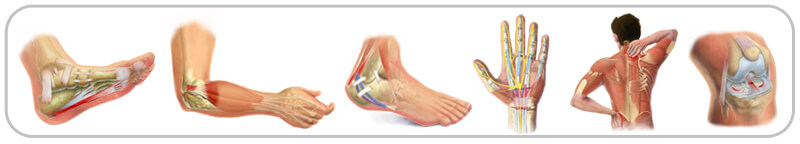
Surgical Treatment
Surgical techniques for rupture repair are varied but usually involve reapproximation of the torn ends of the Achilles tendon, sometimes reinforced by the gastrocsoleus aponeurosis or plantaris tendon. Open reconstruction is undertaken using a medial longitudinal approach. Studies indicate that patients who undergo percutaneous, rather than an open, Achilles tendon rupture repair have a minimal rate of infection but a high rate of sural nerve entrapment (16.7% of treated cases).
Prevention
The following can significantly reduce the risk of Achilles tendon rupture. Adequate stretching and warming up prior to exercising. If playing a seasonal sport, undertake preparatory exercises to build strength and endurance before the sporting season commences. Maintain a healthy body weight. This will reduce the load on the tendon and muscles. Use footwear appropriate for the sport or exercise being undertaken. Exercise within fitness limits and follow a sensible exercise programme. Increase exercise gradually and avoid unfamiliar strenuous exercise. Gradual ?warm down? after exercising.